


Treatments
Pearl testing
The identification of pearls can be challenging in terms of their origin and whether or not theyŌĆÖve been treated. In some cases, there are some simple tests that one can do, but a complete evaluation will normally require examination by a pearl expert, or by the expertise and analytical equipment at a major gem testing laboratory. This is particularly important for certain high-quality natural or cultured pearls that have great value in the market today.
Simple tests
Before submitting pearls to a jeweller or laboratory, there are two very simple tests for basic pearl identification. One technique, known as the ŌĆśtooth testŌĆÖ, can be used to detect an imitation pearl. When rubbed against the edge of a tooth, a natural or cultured pearl will feel gritty or rough, while an imitation pearl will feel very smooth. This test does not help separate the nacreous pearls formed in specific molluscs, though. The other is the ŌĆścandling testŌĆÖ. This works by rotating a cultured pearl in front of a strong light source, such as a penlight. In some directions, a series of parallel lines or bands may be visible, causing a kind of ŌĆśblinkingŌĆÖ effect. These lines correspond to the banding pattern in one plane of the bead nuclei, but for the test to work the nacre covering over the bead has to be very thin so that the banding is visible. If the bands do not show, the pearl is either natural, non-bead cultured, or bead cultured pearl with a nacre overgrowth that is too thick to allow the technique to work.
Advanced tests
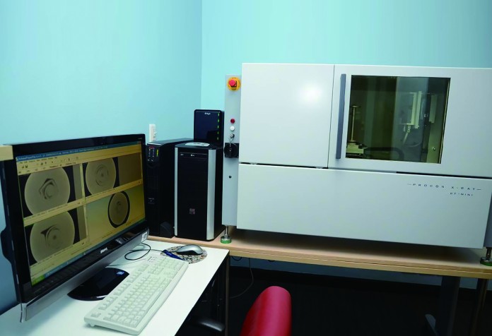
In a gem testing laboratory, pearl identification is based on careful visual examination with a microscope, images of a pearlŌĆÖs internal structure obtained by X-radiography, and analysis of chemical composition and spectroscopy data. Even with the use of these advanced techniques, pearl identification can sometimes still be challenging.
The separation of natural and treated pearls has already been summarised in the preceding sections of this chapter. The principal means for separating natural, bead-cultured, non-bead cultured, composite-cultured, and imitation pearls from one another is the use of X-radiography, which was introduced to gemmology in the 1930s. Although early X-ray machines were somewhat cumbersome to use and potentially unsafe to operate, modern equipment relies on state-of-the-art imaging technology that requires no photographic film or chemicals, and offers a more efficient and economical way of testing.
When X-rays pass through the internal structure of pearls, they are absorbed or transmitted to different extents by the various components that make up the pearl. Those differences are revealed in a photographic film or digital image. Various forms of calcium carbonate form the nacre. The composition of the organic conchiolin, or the presence of voids between nacre layers are depicted as lighter and darker areas of the image, which can be used to identify the natural or cultured origin of a pearl. Pearl testing using X-rays is analogous to the technique used by dentists to check the condition of a personŌĆÖs teeth.
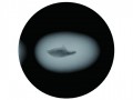
The X-ray patterns of natural pearls typically show a small dark core surrounded by a concentric ring structure. Bead-cultured pearls display an opaque bead surrounded by slightly translucent nacre. Baroque-shaped, bead-cultured pearls, on the other hand, often show darker conchiolin-rich areas and/or voids between the bead and the nacre.
In contrast, the X-radiographs showing the structure of non-bead cultured pearls can be more complicated to interpret. Most show voids that can range in appearance from small linear or twisted structures (freshwater samples) to large cavities of varying outlines (saltwater samples).
When the internal structures of any pearl are difficult to interpret using standard microradiographs, the use of X-ray computed micro-tomography is required. This technique, though costly and time-consuming, produces a 3-D image of the pearl (analogous to a CAT-scan of a human body). Two-dimensional slices through the pearl in any direction may also be obtained from the 3-D model, to provide further assistance in identification. This technique, which is only newly applied in gemmology, is bound to become increasingly important over the years to come.
Other challenges
Composite cultured (including mab├® cultured) and imitation pearls are relatively straightforward to recognise. They can often be identified with the unaided eye, unless they are mounted in bezel settings, which have an open or closed back. A clear boundary usually exists between the cultured pearl face and the mother-of-pearl base. In addition, X-radiography will normally reveal the distinctive structures within the composite. Mab├® cultured pearls are often hollow, and the space contains a filler which can appear on the X-ray images.
Imitation pearls made of glass will show up on X-rays as opaque white spheres, whereas hollow plastic imitation pearls will appear blank on the images. Some solid plastic or shell imitation pearls may well resemble pearls that exhibit little in the way of structure. Since these imitations often have a pearl-like coating on their surfaces, they are not always easy to identify visually.
The experience of the gemmologist, especially using a microscope or other form of magnification to examine pearls, still plays a major role in detecting any type of treatment. Advanced analysis through the aid of a well-equipped gemmological laboratory often serves as a tool for confirming a gemmologistŌĆÖs beliefs, though identification of many treatments requires some type of advanced analysis. While there are always new concerns about ŌĆśundetectableŌĆÖ treatments, researchers are also adapting and adding to the arsenal of sophisticated testing procedures to help them solve such challenges. As identification challenges escalate, gemmologists will continue to work hand-in-hand with technology to resolve them.
TodayŌĆÖs identification challenges have been further complicated by the use of a variety of beads including natural and cultured pearls of all kinds, as well as shells and other materials. These atypical bead cultured pearls have become more abundant in the market, and they can be either very easy or quite difficult to recognise. The advent of such pearls means that traditional microradiographic techniques may not always be sufficient to determine the identity of unknown pearls submitted to laboratories. This is why the use of various CAT X-ray units has become necessary.
(This article is reprinted with permission from GIA in two parts. Part one was published in HKJM DecŌĆÖ13.)
← Back